
Gary Blum, MD
Medical Director, Santa Barbara Office

Jason Barksdale, MD
Radiologist at Pueblo Radiology

Jacob Harter, MD
Radiologist at Pueblo Radiology

John Wrench, MD
Radiologist at Pueblo Radiology

Since their discovery in 1895 by the German physicist Wilhelm Roentgen, X-rays have played a major role in helping physicians diagnose and treat disease. X-rays are high-energy electro-magnetic waves created within an x-ray tube.
They are highly penetrating, and in combination with computer imaging plates, provide images of various internal organs and structures.
Your physician and the radiologist combine to provide you with the test best suited to your particular situation. While all radiation exposure carries some risk, the benefits of diagnosing your condition far outweigh these risks.
However, due to these risks, X-ray examinations are carried out by trained, licensed personnel and interpreted by physicians (radiologists) who are specially trained in the imaging sciences.
The prep for an UGI is found in our appointment prep section. During your exam, the radiologist uses a TV-like x-ray device (fluoroscope) to watch the barium travel down your esophagus (“food pipe”) and into your stomach and small bowel (intestine).
The radiologist will take pictures of the various structures of your GI tract as he/she instructs you to turn from side-to-side.
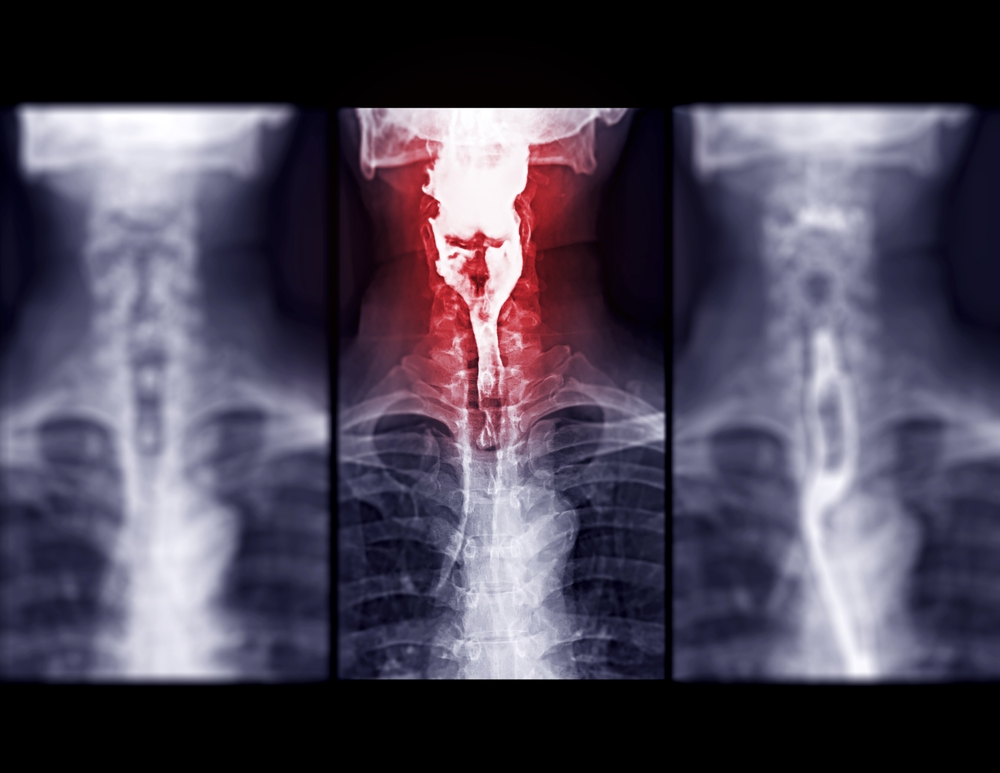
Esophagram or Barium swallow AP view Compare after the patient drinks a liquid that contains barium showing esophagus.
You will be given some crystals and a small amount of water; you will be instructed to swallow the crystals with the aid of the water. Following this, you will be given a cup of barium to drink. The radiologist will instruct you when to drink the barium as he/she watches through a TV-like device. The doctor will examine your swallowing mechanism as well as the various parts of your stomach and take pictures as appropriate. The exam will begin with you standing and then a motorized table will tilt you down to a recumbent position. During the exam, you will be instructed to roll/move to your right and left sides.
The barium will pass through your bowels over the course of a few days. It is important that you drink fluids during this time to help the barium pass through your system. Your stools may be lighter in color for several days.
Barium is a very dense material that allows your GI tract to be visualized with x-rays. In liquid form, barium is the consistency of a thinned milkshake and has no particular flavor. The vast majority of patients tolerate barium well.
Barium studies are typically done in 30 minutes or less.
Barium enemas (BE) are done when there is a suspicion of bowel disease or obstruction. Symptoms of abdominal pain, rectal bleeding, or bloody stools are all indicators for a BE.
Flexible rubber-like tubing is inserted into your rectum. After that, barium fluid is put into your large bowel (intestine) and a radiologist watches the filling through a TV-like x-ray device (fluoroscope). The radiologist will turn you from side to side and gently press on your abdomen. During the filling process, pictures will be taken of the various parts of your large intestine. Once all the necessary images have been acquired, the barium will be drained back into the original barium bag.
The barium will continue to pass in your bowel movements over the course of a few days. It is important that you drink fluids during this time to help the barium pass through your system. Your stools may be lighter in color for several days.
Hysterosalpingography is a test to determine whether a woman’s Fallopian tubes are open, as well as if there is any disease in her uterus. Many times, a hysterosalpingogram is done when a woman is having a difficult time getting pregnant.
Hysterosalpingography unicornuated uterus women radiology
Contrast material (dye that shows up on x-ray) is injected into the uterus through tubing that is inserted through the cervix by the woman’s gynecologist or a Pueblo radiologist. The contrast is viewed with a TV-like device (fluoroscope) while the uterus is filling. Ultimately, in cases of infertility, the goal is to have the contrast material “spill” into the woman’s abdominal cavity, confirming that her fallopian tubes are open (patent).
The contrast material is sterile when injected, and that which spills is naturally absorbed in the abdomen and excreted in the urine. The patient will see no change in urine color or consistency.
When the exam is completed, the catheter is removed and the patient is dismissed. Since residual contrast may continue to seep down from the uterus and you may have some spotting, it is recommended that you wear a pad for a few hours following the test. Normally, no sedation is given for a hysterosalpingogram so you can drive home.
This test is a relatively painless examination.
It will not influence your ability to become pregnant; in fact, it is not uncommon for a woman, who was previously having trouble becoming pregnant, to get pregnant following this test.
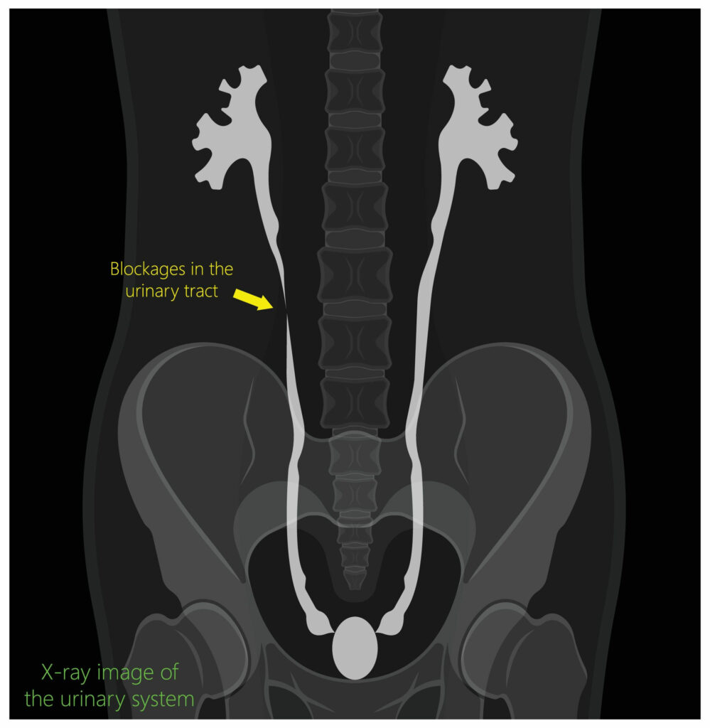
An IVP is a study of the kidneys and bladder and the tubes connecting them called the ureters. An injection of contrast material (dye) is made into a vein in your arm. The contrast material collects and is filtered in your kidneys and ultimately drains down the ureters into your bladder. X-ray films are obtained at various timed intervals following the contrast injection.
It is excreted normally in your urine. As the contrast is mostly clear, you will notice no color change in your urine.
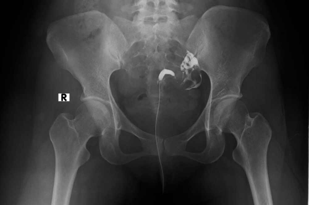
A cystogram is a test where contrast material (dye) is put into your bladder through a catheter (soft, flexible tubing) placed in your urethra. If the urethral catheter is not in place, we will put it in prior to the test. The contrast material will fill your bladder and the radiologist will take pictures of the process. Once the imaging is complete, the dye will be drained out of your bladder.
This is a test of the bladder and the urethra. This test is done when there is a suspicion of disease or abnormality in the mechanism that allows the bladder to empty (voiding / urination). A catheter (soft, flexible tubing) is put into the patient’s bladder through the urethra. The bladder is filled while the radiologist watches the process with a TV-like viewing device (fluoroscope); the radiologist takes pictures at this time. Once the bladder has filled, the catheter is removed and the patient is encouraged to urinate while the radiologist watches and takes pictures of the contrast material moving through the aforementioned structures.

Pueblo Radiology's mission — to provide the highest quality diagnostic imaging in a professional yet compassionate manner — with timely scheduling and results for our patients, referring physicians, and hospital affiliates.
Pueblo Radiology Medical Group
We firmly believe that the internet should be available and accessible to anyone, and are committed to providing a website that is accessible to the widest possible audience, regardless of circumstance and ability.
To fulfill this, we aim to adhere as strictly as possible to the World Wide Web Consortium’s (W3C) Web Content Accessibility Guidelines 2.1 (WCAG 2.1) at the AA level. These guidelines explain how to make web content accessible to people with a wide array of disabilities. Complying with those guidelines helps us ensure that the website is accessible to all people: blind people, people with motor impairments, visual impairment, cognitive disabilities, and more.
This website utilizes various technologies that are meant to make it as accessible as possible at all times. We utilize an accessibility interface that allows persons with specific disabilities to adjust the website’s UI (user interface) and design it to their personal needs.
Additionally, the website utilizes an AI-based application that runs in the background and optimizes its accessibility level constantly. This application remediates the website’s HTML, adapts Its functionality and behavior for screen-readers used by the blind users, and for keyboard functions used by individuals with motor impairments.
If you’ve found a malfunction or have ideas for improvement, we’ll be happy to hear from you. You can reach out to the website’s operators by using the following email
Our website implements the ARIA attributes (Accessible Rich Internet Applications) technique, alongside various different behavioral changes, to ensure blind users visiting with screen-readers are able to read, comprehend, and enjoy the website’s functions. As soon as a user with a screen-reader enters your site, they immediately receive a prompt to enter the Screen-Reader Profile so they can browse and operate your site effectively. Here’s how our website covers some of the most important screen-reader requirements, alongside console screenshots of code examples:
Screen-reader optimization: we run a background process that learns the website’s components from top to bottom, to ensure ongoing compliance even when updating the website. In this process, we provide screen-readers with meaningful data using the ARIA set of attributes. For example, we provide accurate form labels; descriptions for actionable icons (social media icons, search icons, cart icons, etc.); validation guidance for form inputs; element roles such as buttons, menus, modal dialogues (popups), and others. Additionally, the background process scans all of the website’s images and provides an accurate and meaningful image-object-recognition-based description as an ALT (alternate text) tag for images that are not described. It will also extract texts that are embedded within the image, using an OCR (optical character recognition) technology. To turn on screen-reader adjustments at any time, users need only to press the Alt+1 keyboard combination. Screen-reader users also get automatic announcements to turn the Screen-reader mode on as soon as they enter the website.
These adjustments are compatible with all popular screen readers, including JAWS and NVDA.
Keyboard navigation optimization: The background process also adjusts the website’s HTML, and adds various behaviors using JavaScript code to make the website operable by the keyboard. This includes the ability to navigate the website using the Tab and Shift+Tab keys, operate dropdowns with the arrow keys, close them with Esc, trigger buttons and links using the Enter key, navigate between radio and checkbox elements using the arrow keys, and fill them in with the Spacebar or Enter key.Additionally, keyboard users will find quick-navigation and content-skip menus, available at any time by clicking Alt+1, or as the first elements of the site while navigating with the keyboard. The background process also handles triggered popups by moving the keyboard focus towards them as soon as they appear, and not allow the focus drift outside of it.
Users can also use shortcuts such as “M” (menus), “H” (headings), “F” (forms), “B” (buttons), and “G” (graphics) to jump to specific elements.
We aim to support the widest array of browsers and assistive technologies as possible, so our users can choose the best fitting tools for them, with as few limitations as possible. Therefore, we have worked very hard to be able to support all major systems that comprise over 95% of the user market share including Google Chrome, Mozilla Firefox, Apple Safari, Opera and Microsoft Edge, JAWS and NVDA (screen readers), both for Windows and for MAC users.
Despite our very best efforts to allow anybody to adjust the website to their needs, there may still be pages or sections that are not fully accessible, are in the process of becoming accessible, or are lacking an adequate technological solution to make them accessible. Still, we are continually improving our accessibility, adding, updating and improving its options and features, and developing and adopting new technologies. All this is meant to reach the optimal level of accessibility, following technological advancements. For any assistance, please reach out to